Nasal glial heterotopia: A rare interdisciplinary surgical challenge in newborns

Submitted: 2 September 2020
Accepted: 19 March 2021
Published: 20 April 2021
Abstract Views: 4024
PDF: 606
HTML: 14
Publisher's note
All claims expressed in this article are solely those of the authors and do not necessarily represent those of their affiliated organizations, or those of the publisher, the editors and the reviewers. Any product that may be evaluated in this article or claim that may be made by its manufacturer is not guaranteed or endorsed by the publisher.

Authors

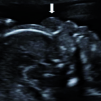
Nasal Glioma (NG) represents a rare congenital abnormality of the neonate, which can be associated with skull defects or even a direct communication to the central nervous system. MRI serves valuable information for differentiation from encephalocele, dermoid cyst and congenital hemangioma. Complete resection remains the treatment of choice. We present two cases of NG, which were both suspected during prenatal ultrasound and MRI. In the first case, postnatal MRI showed a transcranial continuity. Mass excision was performed and the defect was covered by a glabellar flap allowing a good cosmetic result. Postnatal MRI excluded a trans-glabellar communication in the second case. After surgical excision, the resulting skin defect was covered with a full thickness skin graft harvested from the right groin. In cases of NGs complete resection and cosmetic appealing results can be achieved and might necessitate a multidisciplinary approach.


Tirumandas M, Sharma A, Gbenimacho I, et al. Nasal encephaloceles: a review of etiology, pathophysiology, clinical presentations, diagnosis, treatment, and complications. Childs Nerv Syst 2013;29:739-44. DOI: https://doi.org/10.1007/s00381-012-1998-z

Van Wyhe RD, Chamata ES, Hollier LH. Midline craniofacial masses in children. Semin Plast Surg 2016;30:176-80. DOI: https://doi.org/10.1055/s-0036-1593482

Baxter DJ, Shroff MM. Developmental maxillofacial anomalies. Semin Ultrasound CT MR 2011;32:555-68. DOI: https://doi.org/10.1053/j.sult.2011.06.004

Ajose-Popoola O, Lin HW, Silvera VM, et al. Nasal glioma: prenatal diagnosis and multidisciplinary surgical approach. Skull Base Rep 2011;1:83-8. DOI: https://doi.org/10.1055/s-0031-1284210

Rahbar R, Resto VA, Robson CD, et al. Nasal glioma and encephalocele: diagnosis and management. Laryngoscope 2003;113:2069-77. DOI: https://doi.org/10.1097/00005537-200312000-00003

Highton LR, Pay AD. An unusual midline nasal mass in a newborn. J Plast Reconstr Aesthet Surg 2009;62:695-6. DOI: https://doi.org/10.1016/j.bjps.2007.11.064

Hoeger PH, Schaefer H, Ussmueller J, Helmke K. Nasal glioma presenting as capillary haemangioma. Eur J Pediatr 2001;160:84-7. DOI: https://doi.org/10.1007/s004310000602

De Biasio P, Scarso E, Prefumo F, et al. Prenatal diagnosis of a nasal glioma in the mid trimester. Ultrasound Obstet Gynecol 2006;27:571-3. DOI: https://doi.org/10.1002/uog.2754

Penner CR, Thompson L. Nasal glial heterotopia: a clinicopathologic and immunophenotypic analysis of 10 cases with a review of the literature. Ann Diagn Pathol 2003;7:354-9. DOI: https://doi.org/10.1016/j.anndiagpath.2003.09.010

Boyer AC, Krishnan A, Goncalves LF, et al. Prenatal diagnosis of nasal glioma associated with metopic craniosynostosis: case report and review of the literature. J Radiol Case Rep 2015;9:1-8. DOI: https://doi.org/10.3941/jrcr.v9i4.2179

Wassef M, Blei F, Adams D, et al. Vascular anomalies classification: recommendations from the International Society for the Study of Vascular Anomalies. Pediatrics 2015;136:e203-14. DOI: https://doi.org/10.1542/peds.2014-3673

Gasparella, P., Singer, G., Spendel, S., Nagy, E., Eder, H. G., Klaritsch, P., Till, H. ., & Haxhija, E. Q. . (2021). Nasal glial heterotopia: A rare interdisciplinary surgical challenge in newborns. La Pediatria Medica E Chirurgica, 43(1). https://doi.org/10.4081/pmc.2021.240

Downloads

Download data is not yet available.

Citations