Frontal bone fibrous dysplasia in a 6-months-old boy: A distinctive entity
HTML: 15
All claims expressed in this article are solely those of the authors and do not necessarily represent those of their affiliated organizations, or those of the publisher, the editors and the reviewers. Any product that may be evaluated in this article or claim that may be made by its manufacturer is not guaranteed or endorsed by the publisher.
Authors
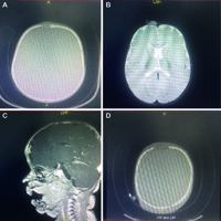
Fibrous Dysplasia (FD) is a non-malignant condition caused by post-zygotic, activating mutations of the GNAS gene that results in inhibition of the differentiation and proliferation of bone-forming stromal cells and leads to the replacement of normal bone and marrow by fibrous tissue and woven bone. The clinical behavior and progression of FD is variable. The management of this condition is difficult and in every case is strictly individualized. We report a case of frontal fibrous dysplasia in a 6month’s old boy who underwent a successfully resection of the lesion with an excellent cosmetic effect.
Altmetrics
Downloads
Citations


How to Cite
PAGEPress has chosen to apply the Creative Commons Attribution NonCommercial 4.0 International License (CC BY-NC 4.0) to all manuscripts to be published.
Similar Articles
- Agostino Berio, Giacomo Garlaschi, Giuseppe Mangiante, Attilia Piazzi, Oculo-auriculo-vertebral spectrum with craniosynostosis and osteo-cartilagineous multiple defects: a diffuse chondro-membranous-osteo-dysplasia , La Pediatria Medica e Chirurgica: Vol. 37 No. 3 (2015)
- Andrea Zangari, Carmine Noviello, Camilla Todesco, Mercedes Romano, Letizia Trotta, Carmine Botta, Ilaria Cascone, Salvatore Scommegna, Gabriele Vasta, Vito Briganti, Alfonso Papparella, Satisfaction and results of the subareolar incision as treatment for gynecomastia in adolescents: experience of two centers , La Pediatria Medica e Chirurgica: Vol. 46 No. 2 (2024)
- Carmine Noviello, Alfonso Papparella, Mirko Bertozzi, Giovanna Riccipetitoni, Ilaria Cascone, Carmine Botta, Giulia Fusi, Veronica Vitali, Mercedes Romano, Abdominal lymphatic malformations in children: case series , La Pediatria Medica e Chirurgica: Vol. 47 No. 1 (2025)
- Agostino Berio, Attilia Piazzi, Carlo Enrico Traverso, Kearns-Sayre syndrome with facial and white matter extensive involvement: a (mitochondrial and nuclear gene related?) neurocristopathy? , La Pediatria Medica e Chirurgica: Vol. 39 No. 4 (2017)
- Muhammad Faizi, Nur Rochmah, Soetjipto Soetjipto, Anang Endaryanto, Sukmawati Basuki, Yuni Hisbiyah, Rayi Kurnia Perwitasari, Protein tyrosine phosphatase non-receptor type 22 C1858T gene polymorphism in children with Down syndrome and autoimmune thyroid diseases , La Pediatria Medica e Chirurgica: Vol. 45 No. 1 (2023)
- Giovanni Murialdo, Attilia Piazzi, Giuseppe Badolati, Enrico Calcagno, Agostino Berio, Oculo-auriculo-vertebral spectrum with myopathy and velopharyngeal insufficiency. A case report with a non-branchiomeric muscle biopsy , La Pediatria Medica e Chirurgica: Vol. 38 No. 2 (2016)
- Gianluca Lista, Fabio Meneghin, Ilia Bresesti, Francesco Cavigioli, Nutritional problems of children with bronchopulmonary dysplasia after hospital discharge , La Pediatria Medica e Chirurgica: Vol. 39 No. 4 (2017)
- Agim Gjikopulli, Sonila Tomori, Marjeta Tanka, Donjeta Bali, Challenges in diagnosis and treatment of Cushing disease in a 12-year-old boy , La Pediatria Medica e Chirurgica: Vol. 47 No. 1 (2025)
- Ilaria Riccio, Elvira Pota, Marco Marcarelli, Maria Carmen Affinita, Daniela Di Pinto, Cristiana Indolfi, Nicola Del Regno, Marco Esposito, Osteonecrosis as a complication in pediatric patients with acute lymphoblastic leukemia , La Pediatria Medica e Chirurgica: Vol. 38 No. 3 (2016)
- Carlo Ripoli, Anna Paola Pinna, Faustina Podda, Roberta Zanni, Maria Giada Tronci, Anna Maria Nurchi, Second-generation antipsychotic and diabetes mellitus in children and adolescents , La Pediatria Medica e Chirurgica: Vol. 39 No. 4 (2017)
You may also start an advanced similarity search for this article.