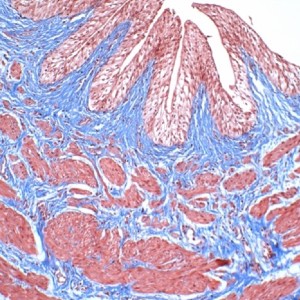
Ureteropelvic junction obstruction in children by polar vessels: histological examination result

Submitted: 17 January 2023
Accepted: 15 May 2023
Published: 30 May 2023
Accepted: 15 May 2023
Abstract Views: 675
PDF: 335
HTML: 8
HTML: 8
Publisher's note
All claims expressed in this article are solely those of the authors and do not necessarily represent those of their affiliated organizations, or those of the publisher, the editors and the reviewers. Any product that may be evaluated in this article or claim that may be made by its manufacturer is not guaranteed or endorsed by the publisher.
All claims expressed in this article are solely those of the authors and do not necessarily represent those of their affiliated organizations, or those of the publisher, the editors and the reviewers. Any product that may be evaluated in this article or claim that may be made by its manufacturer is not guaranteed or endorsed by the publisher.
Similar Articles
- Salvatore Fabio Chiarenza, Elena Carretto, Valeria Bucci, Samuele Ave, Giuseppe Pulin, Cosimo Bleve, Uretero-pelvic junction obstruction in children: Is vascular hitch an effective and safe solutions in very long term outcome? Report of 25 years follow-up , La Pediatria Medica e Chirurgica: Vol. 45 No. 1 (2023)
- Cosimo Bleve, Valeria Bucci, Maria Luisa Conighi, Francesco Battaglino, Lorenzo Costa, Lorella Fasoli, Elisa Zolpi, Salvatore Fabio Chiarenza, Horseshoe kidney and uretero-pelvic-junction obstruction in a pediatric patient. Laparoscopic vascular hitch: A valid alternative to dismembered pyeloplasty? , La Pediatria Medica e Chirurgica: Vol. 39 No. 4 (2017)
- A. Marte, M. Prezioso, L. Pintozzi, S. Cavaiuolo, S. Coppola, M. Borrelli, P. Parmeggiani, Laparoscopic treatment of UPJ obstruction in ectopic pelvic kidneys in children , La Pediatria Medica e Chirurgica: Vol. 34 No. 5 (2012)
- George Vlad Isac, Gabriela Mariana Danila, Sebastian Nicolae Ionescu, Spontaneous resolution and the role of endoscopic surgery in the treatment of primary obstructive megaureter: a review of the literature , La Pediatria Medica e Chirurgica: Vol. 45 No. 2 (2023)
- Salvatore Fabio Chiarenza, Cosimo Bleve, Ciro Esposito, Maria Escolino, Fabio Beretta, Maurizio Cheli, Vincenzo Di Benedetto, Maria Grazia Scuderi, Giovanni Casadio, Maurizio Marzaro, Leon Francesco Fascetti, Pietro Bagolan, Claudio Vella, Maria Luisa Conighi, Daniela Codric, Simona Nappo, Paolo Caione, Guidelines of the Italian Society of Videosurgery in Infancy for the minimally invasive treatment of the ureteropelvic-junction obstruction , La Pediatria Medica e Chirurgica: Vol. 39 No. 3 (2017)
- Rossella Angotti, Giulia Fusi, Elena Coradello, Clelia Miracco, Francesco Ferrara, Marina Sica, Alessandra Taddei, Gabriele Vasta, Mario Messina, Francesco Molinaro, Lichen sclerosus in pediatric age: A new disease or unknown pathology? Experience of single centre and state of art in literature , La Pediatria Medica e Chirurgica: Vol. 44 No. 1 (2022)
- Marco Gasparella, Maurizio Marzaro, Mario Ferro, Carlo Benetton, Vittorina Ghirardo, Cinzia Zanatta, Francesco Zoppellaro, Meckel’s diverticulum and bowel obstruction due to phytobezoar: a case report , La Pediatria Medica e Chirurgica: Vol. 38 No. 2 (2016)
- M. Gasparella, M. Ferro, M. Marzaro, C. Benetton, C. Zanatta, F. Zoppellaro, Acute abdomen in children: a continuous challenge. Two cases report: Meckel’s Diverticulum with Small Bowel Volvolus and Internal Herniation related to Epiploic Appendagitis mimicking acute appendicitis , La Pediatria Medica e Chirurgica: Vol. 36 No. 2 (2014)
- A. Marte, L. Pintozzi, M. Prezioso, M. Borrelli, S. Pisano, Psychopathologic risk assessment and self-esteem in patients undergoing hypospadias surgery , La Pediatria Medica e Chirurgica: Vol. 36 No. 2 (2014)
- Arianna Mariotto, Nicola Zampieri, Mariangela Cecchetto, Francesco Saverio Camoglio, Ureteral rupture after blunt abdominal trauma in a child with unknown horseshoe kidney , La Pediatria Medica e Chirurgica: Vol. 37 No. 2 (2015)
You may also start an advanced similarity search for this article.

 https://doi.org/10.4081/pmc.2023.308


